
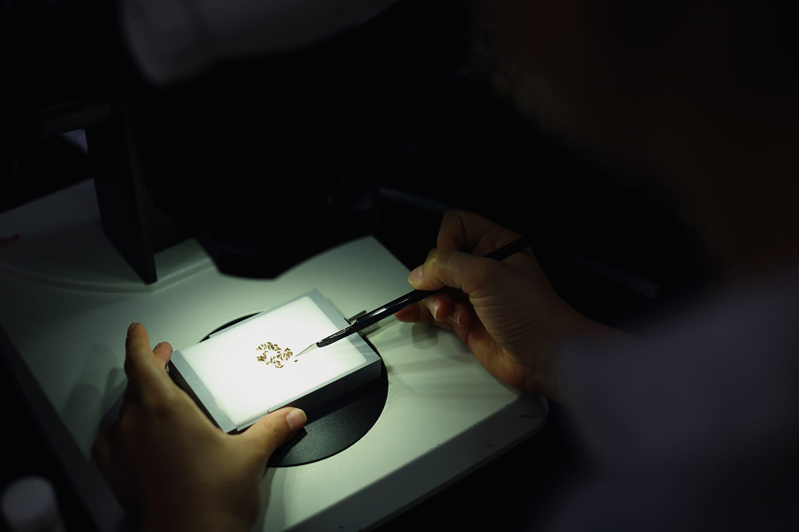
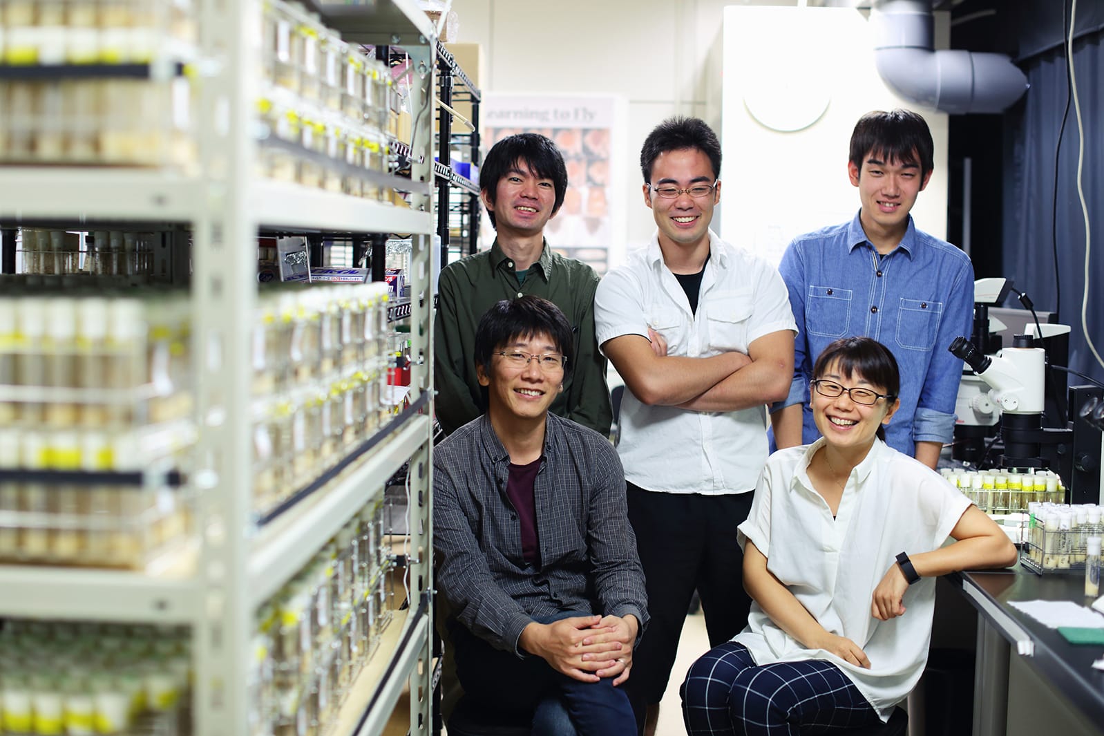
As multicellular organisms, we get our shape from an astounding process that starts with a single cell in the form of a fertilized egg. For example, the human body is formed from a collection of over 60 trillion individual cells. However, just as you cannot build a building simply by stacking bricks, you cannot form a body simply by putting cells together. “Cells” only become “an organism” when a disorderly clump of cells becomes ordered. To unravel the mysteries of this process, we focus on the process by which cells begin to shape the body. Now I investigate the fruit fly Drosophila, a small animal suited to research that we are using as a model animal. This insect likes natural fruit juice, so it’s called “fruit fly”. It’s about 2 mm in body length and is bred in palm size. As an animal that undergoes complete metamorphosis, Drosophila undergoes dynamic body shaping when it transforms from a rod-shaped larva to an adult with a head, thorax, and abdomen during the pupal stage. Although such dramatic changes are occurring, live imaging of organogenesis is very easy because the flies do not drink, eat, or move during the pupal stage. We perform the live-imaging of organogenesis under a laser microscope while the pupae are still alive. Using Drosophila genetics, we induce expression of fluorescent proteins such as GFP in the cells or tissues we want to study. We can then visualize cells, organelles, and proteins and analyze the dynamics that govern cooperation among cell groups.

Those “study” and “idea” become the trigger of “to investigate”.
- Erina KURANAGA
- Professor of department of Biology, Faculty of Science and graduate school of Life Sciences
1.
What kind of research are you doing?
2.
What is the reason for starting your study?
When I faced the fact that a beautiful swallowtail butterfly was originally a caterpillar, I was shocked while I was a child, and I could not help feeling the mystery of life. As I grew up, I began to think that the events happening in the pupa may be the same as what is happening in the uterus of our mother, first of all I would like to understand about small insects.
3.
Message for prospective students
We are surrounded by many wonders and mysteries. Please do not settle with the word just “mystery”, first study it and consider by yourself. Those "study" and "idea" become the trigger of "to investigate". And if you want to investigate more, please come to Tohoku University's Faculty of Science, and why don’t you “research” the mystery with us?
- Name:
- Erina KURANAGA
- Position:
- Professor of department of Biology, Faculty of Science and graduate school of Life Sciences
- Laboratory:
- Histogenetic Dynamics
- Hometown:
- Kumamoto
- Research area:
- Classical and modern mystery novels (but recently I have read “Godzilla GENRON” by Shigeru Kuratani)
- Posted Date:
- Jan 24, 2018
JAEN
